WHAT IS ULTRASOUND IMAGING OF THE PELVIS?

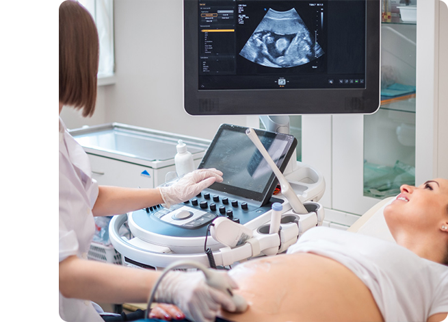
Ultrasound imaging involves exposing parts of the body to a high frequency sound wave in order to produce images of the uterus, ovaries, fallopian tubes and other surrounding structures. It is a non invasive medical test that may help your physician or health care provider diagnose certain medical conditions.


There are two types of pelvic ultrasounds:

  • Trans-Abdominal(images taken from the surface of the abdomen)
  • Trans-vaginal(images taken with an ultrasound probe inside of the vagina)

Both of these tests work well in combination and can be highly diagnostic.

Preparation

A full bladder is required for a pelvic ultrasound. Patient’s should drink 1 litre of fluid one hour prior to the examination and ensure to not empty your bladder prior to the ultrasound. Once the ultrasound is complete you will be able to void.

Transvaginal

Transvaginal ultrasound is performed in a similar way to a gynaecological examination (patient lying on her back with feet in stirrups) and is completed with an empty bladder. The ultrasound probe is covered with a disposable non-latex protective sheath after following all guidelines for high level disinfection/sterilization. A small amount of lubricant is placed on the tip of the probe and it is then gently inserted into the vagina.

Antenatal-care
image-layers-3_2
image-layers-3_3
 
The images are obtained from different orientations in order to get detailed images of the uterus and ovaries. Please let your sonographer know if you experience any pain or discomfort. It is common for some minor pressure or tenderness to be felt when scanning.